Nearly all primary care providers in the United States, especially those with sizeable HIV practices, are aware of the very real dangers of crystal methamphetamine use. It has been a pervasive concern among many Physicians’ Research Network members—indeed, a hot topic during several question-and-answer sessions—and an issue that PRN has been struggling to adequately address.
PRN’s first agenda item was to find a speaker to lay out some of the potential physiologic concerns of crystal meth use among HIV-positive people. We were promptly guided to Dr. Scott Letendre of the University of California, San Diego, who has been engaged in some significant research involving the effects of methamphetamine on central nervous system function in HIV-positive people. We present here a summary of his June 2005 lecture and intend to publish additional articles in coming Notebook issues, to increase awareness among clinicians regarding the very real dangers of methamphetamine addiction and its potential management.
| I. Crystal Methamphetamine: An Introduction | Top of page |
The ease with which crystal meth can be manufactured is a major contributing factor to the increase in its use. Law enforcement officials identify and close hundreds of clandestine methamphetamine labs each year. Large operations produce methamphetamine in Mexico and California. Outside of these areas, small rural laboratories are more common. Rural areas are favored because strong odors are produced in the process of “cooking” the drug, which can draw immediate attention to the manufacturing operation.
The manufacturing of crystal meth begins with large quantities of ephedrine or pseudoephedrine, which are found in numerous over-the-counter cold medications (manufacturers of these compounds are currently looking into producing pseudoephedrine-free products). These are then cooked with a variety of other readily available substances, including red phosphorus, hydrochloric acid, drain cleaner, battery acid, lye, paint thinner, Freon, and/or antifreeze (every pound of crystal meth produced leaves behind five to six pounds of toxic waste). And because it is so easily and rapidly produced, it remains one of the cheapest drugs available.
With the manufacturing process completed, crystal meth is bagged and sold as shiny yellowish-white rocks in various sizes or as a crystalline powder resembling small fragments of glass. It can be ingested by snorting or smoking, dissolved in water to be swallowed or inserted into the rectum (“booty bump”), or injected intramuscularly or intravenously. On the street, it goes by a variety of names, including crystal, glass, crank, Tina, Crissy, and ice (in its rock form).
Figure 1.Methamphetamine Administration: Geographic Variations

The preferred method of taking methamphetamine varies among geographic regions. The bar graph is based on surveys conducted by the National Institute of Drug Abuse Community Epidemiology Work Group in three U.S. regions: San Diego (survey conducted between July and December 2000), Minneapolis/St. Paul (survey conducted throughout 2000), and Texas (survey conducted in January through June 2001).
Source: NIDA Community Epidemiology Work Group
“The preferred route of administration varies by geographical region,” Dr. Letendre commented (see Figure 1). “In San Diego, people have learned that smoking meth gets the drug into the system quickly. In Texas, the preferred route is intravenous administration. In New York, the preferred route seems to be intravenous injection as well.” According to the National Institute of Drug Abuse (NIDA), the bioavailability of methamphetamine administered via the oral route is 67.2%. Through the intranasal route, the bioavailability is 79%. Smoking it increases the bioavailability to 90.3% and injecting it intravenously increases the bioavailability to 100%
Plasma concentrations of methamphetamine peak approximately 15 minutes after it is administered. Concentrations drop by approximately 50% within 30 minutes, followed by steady-state concentrations for another 90 minutes or so. “However, if we follow the concentration curve, we see that the drug remains in the system for more than 24 hours,” Dr. Letendre explained. “Metabolites in the urine can be detected out to three or four days.”
Methamphetamine is hepatically metabolized via deamination, conjugation, and hydroxylation. It is metabolized to amphetamine through cytochrome P450 2D6. “One potential concern here is the effect of genetic variation in 2D6 on methamphetamine concentrations,” Dr. Letendre commented. “Such genetic variations can result in poor and extensive metabolizer phenotypes.” Also of concern is the fact that ritonavir (Norvir) inhibits 2D6 and can result in threefold to tenfold increases in methamphetamine concentrations. “Several deaths have been reported from possible methamphetamine-protease inhibitor interactions,” he warned.
| Methamphetamine and Biological Markers | Top of page |
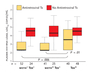
Dr. Letendre’s group has also evaluated plasma and CSF HIV-RNA levels in HIV-positive individuals with a history of methamphetamine use (Ellis, 2003). Three groups were evaluated: 88 subjects with a history of methamphetamine use with evidence of recent use, determined by positive toxicology results (meth+/tox+); 66 subjects with a history of methamphetamine use with no evidence of recent use (meth+/tox-); and 76 subjects without a history of methamphetamine use and no evidence of recent use (meth-/tox-). More than half of the subjects were receiving antiretroviral therapy at the time of study entry.
Figure 3.Relationship of Antiretroviral Therapy to Plasma HIV-RNA Levels in Three Study Groups

Box-and-whisker plots show the median (center line), interquartile range (box) and 5th and 95th percentiles (whiskers) for subjects receiving antiretroviral therapy and for those not receiving antiretroviral therapy. Meth-/tox-: no history of methamphetamine dependence or current use; meth+/tox-: history of methamphetamine use but no current use; meth+/tox+: history of methamphetamine use and evidence of recent use.
Source: Ellis, 2003. The Journal of Infectious Diseases 188:1822. Adapted with permission of the Infectious Diseases Society of America.
The meth+/tox+ patients had plasma HIV-RNA levels that were significantly higher than either the meth+/tox- or meth-/tox- patients. However, adjustment for antiretroviral use identified that the only significant differences were among the subjects receiving treatment (see Figure 3). Compared to subjects receiving antiretroviral therapy in the meth+/tox- and meth-/tox- groups, subjects receiving antiretroviral therapy in the meth+/tox+ had significantly higher viral loads. In contrast, HIV-RNA levels among meth+/tox+ subjects not receiving antiretroviral therapy were not significantly higher than those in the meth+/tox- or meth-/tox- groups. This, Dr. Letendre suggested, argues against a direct biological effect of methamphetamine use itself on viral replication. Rather, it suggests that recent methamphetamine use and antiretroviral therapy interact in their effects on viral load. This, he said, “is no less of a significant problem.”
No evidence of increased CSF viral load in meth+/tox+ patients was documented, compared to meth+/tox- or meth-/tox- study volunteers.
The increases in plasma viral load found among the meth+/tox+ subjects receiving antiretroviral therapy may be due to poor adherence or to altered metabolism of antiretroviral medications. This study, however, was unable to determine the exact cause. Dr. Letendre’s group collected self-reports of adherence and found them to be similar in the meth+/tox+ subjects, compared with subjects in the other groups. However, he noted, self-reports of adherence to therapy can be inaccurate.
Dr. Letendre also pointed out that, at baseline, the duration of methamphetamine abstinence correlated with HIV-RNA levels in plasma and in CSF. And in the longitudinal analysis, relapsing methamphetamine users had higher HIV-RNA levels in plasma (4.4 log10 copies/mL vs. 2.3 log10 copies/mL) and in CSF (2.4 log10 copies/mL vs. 1.7 log10 copies/mL) compared with those who remained abstinent.
Additional data from UCSD indicate that methamphetamine use is associated with increased inflammation in HIV-positive individuals. Dr. Letendre explained that HIV-positive methamphetamine users had higher levels of five markers of macrophage activation—including MCP-1, sCD14, sTNFR-II, TNF-a, and MIP-1 beta—in plasma. Three markers were also higher in the CSF of methamphetamine users. And similar to HIV-RNA levels, macrophage activation levels varied with recency of methamphetamine use.
| Methamphetamine and Neurobiology in HIV | Top of page |
Dr. Eliezer Masliah, Professor of Neuroscience and Pathology at UCSD, and his colleagues have been conducting a great deal of neurobiological research surrounding the effects of methamphetamine in the brains of HIV-positive individuals. His team’s research has focused on tissue samples collected at autopsy, which includes deceased individuals enrolled in the UCSD program project and samples retrieved from the National NeuroAIDS Tissue Consortium, which includes sites in San Diego, New York, Los Angeles, and Galveston.
To date, Dr. Masliah’s group has analyzed data from human brain tissue studies, animal studies, and in vitro experiments. Dr. Letendre reported on the human studies that used brain tissue from 19 HIV-negative volunteers (controls), some of whom had a history of methamphetamine use; 42 HIV-positive people with no history of methamphetamine use, and 27 HIV-positive people with a history of methamphetamine use (Langford, 2003). Samples were collected from patients who died around 40 years of age. A number of the HIV-positive patients had evidence of HIV encephalopathy at the time of death.
Overall, HIV-positive meth users more frequently had evidence of ischemic events, severe inflammation (as measured by microglial reactivity), and loss of synapses (as measured by synaptophysin) than HIV-positive people who did not use meth. While neuronal and dendritic structures were preserved in the control individuals, meth users who did not have HIV encephalitis (HIVE-negative) had moderate neuronal and dendritic damage, non-meth users who had HIVE had moderate-to-severe damage, and individuals who had both HIVE and meth use had the most severe damage. This last group also had extensive loss of calbindin-immunoreactive interneurons. This is important because loss of these interneurons interferes with neurotransmission of other neurons and has been implicated in other neurologic disorders, such as schizophrenia (Eyles, 2002) and motor neuron disease (Maekawa, 2004). “We have neuropsychological test scores for these patients, conducted before their death and the donation of brain tissue,” Dr. Letendre said. “Dr. Masliah’s group found that greater loss of interneurons among the HIV-positive methamphetamine users correlated with worse neuropsychological performance prior to death.”
These data—along with those being generated by ongoing studies—indicate that methamphetamine and HIV independently injure neural cells, such as microglia, and neurons by several mechanisms, leading to impaired cognition among HIV-positive methamphetamine users.
| References | Top of page |
